Kohn Lab

The University of Michigan School of Dentistry

LEARN MORE

ABOUT

Biomimetics and Biomineralization Research

kohn lab structure function relations

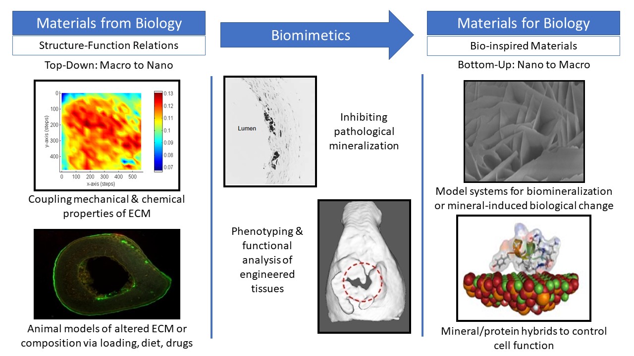
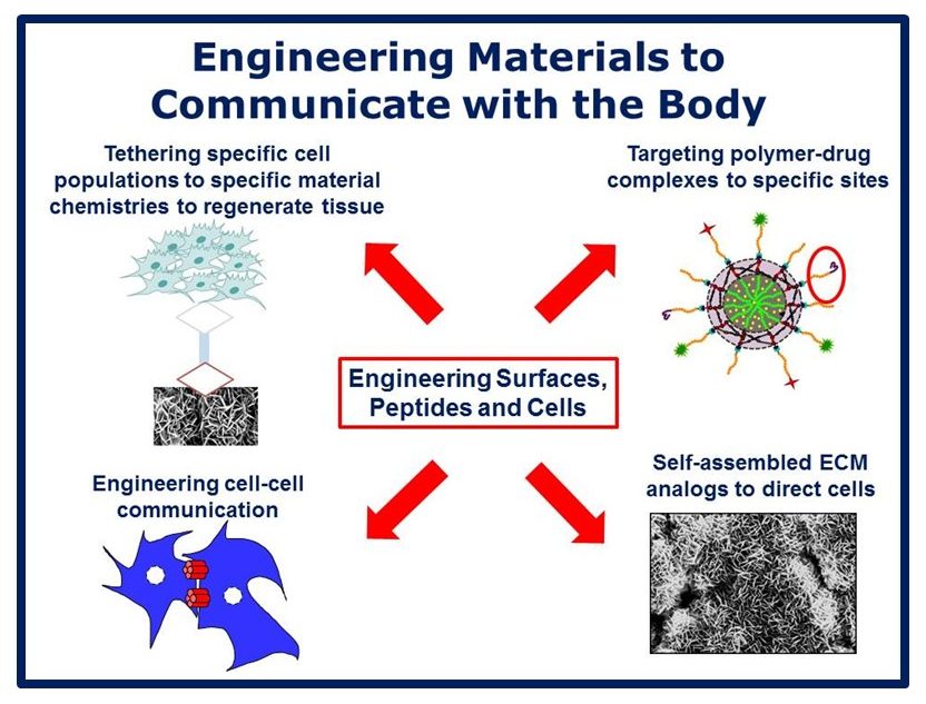
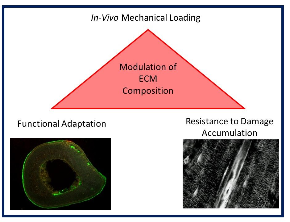
Musculoskeletal disorders incur more health care costs than breast, lung and prostate cancer combined and consume 6% of the US gross domestic product (US Bone & Joint Initiative: The Burden of Musculoskeletal Diseases in the US, 2018). Osteoporotic fractures are responsible for more hospitalizations in the US than heart attacks, strokes, and breast cancer combined (National Osteoporosis Foundation, 2019). Our laboratory is dedicated to understanding causes of skeletal fragility and developing technologies to regenerate skeletal tissue. We investigate mineralized tissues across many levels of scale to establish structure-function relations across their hierarchy and utilize this information to develop biomimetic strategies to regenerate tissues. By coupling mechanical, compositional and molecular analyses of tissues, we investigate mechanisms of skeletal fragility and mechanically mediated tissue adaptation. Using principles of biomineralization, we design materials that can communicate with their biological microenvironment, leading to better control of cell function in-vitro and tissue formation in-vivo. Our work has been funded from the NIH, NSF, DoD, and industry. We have published over 145 peer-reviewed papers and book chapters, hold 10 patents, and have given over 130 invited presentations. Dr. Kohn has mentored 45 graduate students, 7 post docs, 50 undergraduates, 14 residents and 5 visiting professors in his laboratory and many past trainees are now funded independent investigators.

PROJECTS

Biomimetic Biomaterials

A limitation in translating tissue engineering to the clinic is an inability to reproducibly achieve a significant volume of tissue. To better control cells’ response to biomaterials and guide tissue regeneration, we mimic aspects of nature’s biomineralization strategies as a basis for biomaterials design. We developed biomaterials based on the self-assembly of biological mineral onto 3D organic templates. We also demonstrated that the function of stem cells and amount and spatial distribution of bone regenerated in-vivo were enhanced by controlling the nucleation and growth of bioimimetic mineral onto an organic template. The combination of material chemistry and substrate rigidity provide substrate-mediated cues for control of cell behavior, but soluble signals from a biomaterial can also influence cell differentiation, demonstrating that a biomaterial can influence cells without being in direct contact. This finding led us to engineer cell-cell communication as a strategy to enhance regeneration. We have shown that alterations in cell-cell communication via overexpressing gap junction proteins leads to significantly greater volumes of bone formed in-vivo and more uniform spatial distribution, potentially overcoming a critical barrier in tissue engineering.

biomimetic biomaterials image

Biomaterials that Communicate with the Biological Milieu

We have developed organic/inorganic hybrid biomaterials in which proteins direct biological response to the material either passively or actively. The former technique capitalizes on Nature’s mechanisms of controlling crystal growth via protein/crystal binding, while in the later technique, therapeutic agents are integrated into the mineral in a spatially controlled manner, allowing for spatial and temporal delivery of active biological molecules. We developed high throughout combinatorial approaches to discover novel peptides and investigate peptide/biomaterial interactions, Using phage display, we discovered peptides that exhibit preferential affinity to specific material chemistries and a second set of peptides with affinity to specific cells. We combined these sequences to create dual-functioning peptides that have material and cell-specific binding domains, serving as tethers between the material and biological worlds and enabling spatially directed tissue formation. Capitalizing on the peptides’ apatite-binding binding ability, we next demonstrated their ability to bind to cell-secreted mineral, and these peptides are now used to inhibit pathological calcification and bind to bone for targeted delivery of drugs.

biological milieu image

Structure-Function Relations in Mineralized Tissues Across Hiearchy

To develop structure-function relations in mineralized tissues and define metrics for bone quality across dimensional scale, we have used acoustic emission, Raman spectroscopy, NMR and HPLC to detect and predict damage, and identify ultrastructural changes associated with compromised bone quality. In collaboration with Dr. Michael Morris, we were the first to couple mechanical and compositional analyses of tissues using Raman spectroscopy, and relate mechanical deformation at the ultrastructural level to compositional changes. Using NMR, we are amongst the first to identify the important role of water in mediating mechanical stability of bone, and the first to demonstrate that there are 3 distinct water compartments in bone, each mediating a different functional response. Most recently, we developed a new animal model of lathyrism and HPLC methods for the direct quantification of immature and mature cross-links. We identified significant positive correlations between pyridinoline cross-link content and tissue strength and we are the first to report a correlation between bone fracture toughness and cross-link maturity. A significant outcome of this work is the recognition that a critical determinant of bone quality is cross-link type, rather than total quantity, and the cross-links that exist in lower abundance are the best predictors of bone quality.

minerilized tissue image

Functional Adaptation in Bone

Building on our ex-vivo based structure-function findings, we developed a short-term, weight-bearing rodent exercise model. Using this model with mice of different ages, genders, background strains and genetic modifications, we identified significant location and gender-specific differences in phenotype and mechanical responsiveness of the skeletal system. We also determined that exercise can rescue deficiencies in function caused by gene deletion, suggesting that mechanical stimulation can compensate for genetically-induced deficiencies in bone quality. However, we found that the phenotype resulting from targeted deletion of bone matrix genes is dependent on the background strain on which transgenic mice are bred. Using the exercise model, we also demonstrated that in-vivo mechanical loading can increase resistance to fatigue damage, and defined compositional changes in collagen cross-linking guiding the increased resistance. Most recently, using exercise in conjunction with enhancement or inhibition of PTH signaling, we demonstrated that bone adaptation during exercise is not only a function of dynamic loading, but also PTH release, and that PTH signaling contributes differently to bone adaptation at the structural and tissue levels.

functional adaptation image

How Mechanical Competence of Bone can be Explained by Compositional Changes

It has long been assumed that bone adapts to mechanical loading and improves its strength by turning over or adding material. We demonstrated that bone can also respond to exercise, as well as genetic alterations and other stimuli/insults, via mechanical changes that are not mediated by the addition of new bone, but via compositional changes. We have further demonstrated that these compositional changes occur predominantly in the perilacunar zone around osteocytes. We also demonstrated that exercise can shift cross-link chemistry toward the pyridinoline pathway and counter the effects of cross-link inhibition on mechanical properties. Also of significance is our finding that cross-link inhibition and rescue can result in changes in modulus without changes in mineral density, challenging the tenet that modulus is controlled by the mineral component of bone.

mechanical competence image

DIRECTOR

david kohn

David H. Kohn, MSE, PhD
Professor

dhkohn@umich.edu

Dr. Kohn is the Natalie C. Roberts Endowed Professor at the University of Michigan with appointments in the Departments of Biomedical Engineering (College of Engineering) and Biologic & Materials Sciences (School of Dentistry). He received his BS in biomedical engineering from Tulane University and his Ph.D. in bioengineering from the University of Pennsylvania. Dr. Kohn is the PI and Director of 3 large institutional programs in regenerative medicine – an NIH Training Program in Tissue Engineering and Regeneration; the Michigan-Pittsburgh-Wyss Regenerative Medicine Resource Center, an NIH-supported national center whose mission is to catalyze the translation of tissue engineering technologies to the clinic and private sector; and a University of Michigan Biosciences Initiative in Programmable Biomaterials for Regenerative Medicine. Dr. Kohn has served as Graduate Chair in Biomedical Engineering, and was a Visiting Professor in the Craniofacial and Skeletal Diseases Branch of the NIH intramural laboratories. He also serves on the advisory boards of several universities and has consulted for many medical device companies. Dr. Kohn is a Past President of the Society for Biomaterials.

Dr. Kohn has been the recipient of a Whitaker Foundation Biomedical Research Award, NSF Research Initiation Award and NIH IPA award (Visiting Professor at NIH). He is a Fellow of the American Institute of Medical and Biological Engineering (AIMBE), AAAS and the International Union of Biomaterials Scientists and Engineers, and a recipient of an IADR Distinguished Scientist Award.

PUBLICATIONS


View the complete listing of publications.

CONTACT

Kohn Lab
University of Michigan School of Dentistry
1011 N University Ave,
Room D2213
Ann Arbor, MI 48109
Phone: (734) 764-2206

https://kohnlab.bme.umich.edu